
Patella Instability & MPFL Reconstruction in Reading — Private Knee Surgery
Patella instability occurs when the kneecap (patella) slips out of its groove, often after an initial dislocation. This can lead to pain, swelling, and a sense of the knee "giving way." Repeated dislocations may damage cartilage and bone, causing long-term problems if untreated.
Mr Sam Nahas, consultant knee surgeon at Circle Reading Hospital, specialises in the surgical management of recurrent patella instability using MPFL reconstruction, bioabsorbable fixation for osteochondral fractures, and tibial tuberosity realignment where indicated.
Patients benefit from self-pay consultations within a week, rapid MRI access, and comprehensive evaluation of all causes of instability — from soft-tissue injury to underlying bone or rotational alignment issues.
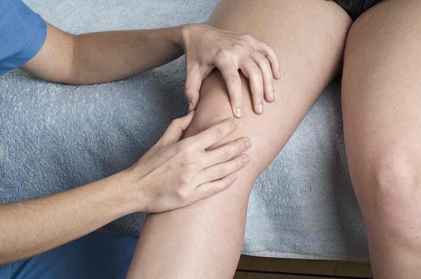
Understanding patella instability
The patella sits within a groove at the front of the femur called the trochlea. The medial patellofemoral ligament (MPFL) is the main structure that prevents the kneecap from dislocating laterally (outwards).
When this ligament tears — usually during a twisting injury or fall — the patella can slip out repeatedly, especially during sport or knee bending.
Common symptoms include:
Feeling of the knee "slipping out" or giving way
Pain and swelling after dislocation
Difficulty trusting the knee during stairs or sport
Visible deformity or bruising after an acute episode
Diagnosis and imaging
Diagnosis is made through a combination of clinical examination and imaging.
MRI helps confirm an MPFL tear and assess associated cartilage or bone injuries
CT scan may be used to evaluate patella alignment and the tibial tuberosity position (TT-TG distance) or rotation of the leg
X-rays identify bony avulsion or trochlear dysplasia (shallow groove)
Patients typically receive MRI within a week and tailored treatment plans within a week.
Surgical options
Treatment depends on the underlying cause and frequency of instability.
MPFL Reconstruction
The most common surgical treatment for recurrent instability is MPFL reconstruction.
Mr Nahas performs this using a hamstring graft (usually from the gracilis tendon) to recreate the torn ligament and restore stability. The graft is anchored using anchors (special screws) in the kneecap and a screw in the femur (thigh).
Osteochondral Fracture Fixation
In first-time dislocations, a small fragment of bone and cartilage may break off (osteochondral fracture).
Mr Nahas fixes these fragments using bioabsorbable screws, restoring smooth cartilage and protecting the joint from early arthritis.
Tibial Tuberosity Realignment
If the patella is being pulled laterally due to an abnormal bone alignment, a tibial tuberosity transfer (realignment) may be performed.
This repositions the patella tendon attachment on the shin bone to improve tracking and stability, often combined with MPFL reconstruction for best results.
When Surgery May Not Be Appropriate under Mr Nahas
In some cases, patella instability results from rotational bone abnormalities or severe trochlear dysplasia (shallow groove). Mr Nahas does not perform these complex bony corrections but will ensure timely referral to trusted colleagues with subspecialty expertise in these conditions.
Recovery and rehabilitation
Recovery after MPFL reconstruction or tibial tuberosity realignment follows a structured pathway:
Weight bearing: may be partial for 4 weeks, progressing with physiotherapy, but depends on the extent of the procedure.
Range of motion: depends on the procedure undertaken by may be restricted early on after a bony procedure
Return to driving: around 6 weeks, once you can perform an emergency stop safely and are off strong painkillers; this also depends on which leg was operated on and whether you drive a manual (see Driving after knee surgery)
Return to sport: 6–9 months, depending on progress and strength
Each patient receives a personalised rehabilitation plan to restore confidence, strength, and stability. These timelines are not guaranteed and can vary between patents.
Evidence and outcomes
MPFL reconstruction is a keyhole operation that rebuilds the main ligament stopping the kneecap from popping out; in people with repeat dislocations—or who are high-risk after a first dislocation—it greatly cuts the chance of it happening again and improves knee function (across 19 studies, early surgery had ~7% redislocation vs ~30% with rehab alone) (Cohen et al., KSSTA 2022).
Randomized and broader systematic reviews show surgery reduces redislocation versus non-operative care—e.g., a meta-analysis of 10 RCTs favored surgery for fewer recurrences and better function, and a KSSTA review reported ~7% redislocation after early MPFL reconstruction versus ~30% with rehabilitation—while in the ≥5-year systematic review and meta-analysis that Mr Nahas co-authored (Cureus 2025), the pooled redislocation rate was 5.75% with a 0.38% reoperation rate alongside consistently favorable patient-reported outcomes, supporting durable benefit of MPFL reconstruction (Xing et al., 2020; Cohen et al., 2022; Cureus 2025).
Why choose Mr Sam Nahas
Consultant knee-only surgeon specialising in patella and soft-tissue reconstruction
Expertise in MPFL reconstruction, tibial tuberosity realignment, and bioabsorbable fixation
Evidence-based practice guided by current BOA and ESSKA standards
Rapid diagnosis and clear, honest discussion of all treatment options
Trusted by physiotherapists and sports clinicians across Berkshire
Self-pay and appointments
Self-pay consultations are available with access to MRI within a week and surgical planning. No GP referral is required.
Location
All procedures are performed at Circle Reading Hospital, serving patients from Reading, Berkshire, Wokingham, Henley, Oxford, and surrounding areas.